Sunday, 16 December 2018

Rehabilitative Exercises for Biceps Tendinitis

Biceps Tendonitis and its exercises

Biceps Tendonitis

Biceps tendonitis is inflammation of the tendon around the long head of the biceps muscle. Biceps tendinosis is caused by degeneration of the tendon from athletics requiring overhead motion or from the normal aging process.


Symptoms of Bicipital Tendonitis

Pain in moving the upper arm is a common symptoms of bicipital tendonitis.
Tenderness and pain in the shoulder
Snapping sound in the shoulder
         Difficulty in lifting something, condition worsening with lifting.

Weakness

Feeling of warmth in the shoulder region

Redness.

Epidemiology of Bicipital Tendonitis


Bicipital Tendonitis is common amongst athletes, especially those, who are associated with sports like tennis, cricket, baseball, weightlifting, kayaking etc. In fact, wheelchair athletes, due to excessive use of hand movements can also have inflammation of the tendons causing the condition. It is also seen in older patients, where the condition is caused by degenerative tendinosis.

Causes of Bicipital Tendonitis


Overuse, repetitive and continuous usage of the biceps tendon can cause Bicipital tendonitis. Due to overuse, damaged cells of the tendons fail to repair themselves. The lack of time to recuperate leads to inflammation and causing bicipital tendonitis. The common causes of Bicipital Tendonitis are –
  • Repetitive sports activities and frequent work activities. Athletes, who swim, throw or use racquets and swing, are mostly affected by bicipital tendonitis.

  • If the shoulders are torn and wearied for years and have not had the time to heal, it can lead to bicipital tendonitis.

  • When the collagen arrangement of the biceps tendons is disrupted, it can lead to inflammation of the tendons.
Rehabilitative Exercises

You may do these exercises right away. If any exercise increases your pain, stop doing it. Avoid overhead lifting while your tendon is healing. Some of most useful and valuable exercises to get recovery in biceps tendinitis are

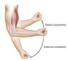
Active elbow flexion and extension: Gently bring the palm of the hand on your injured side up toward your shoulder, bending your elbow as much as you can. Then straighten your elbow as far as you can. Repeat 15 times. Do 2 sets of 15

Biceps stretch:
Stand facing a wall (about 6 inches, or 15 centimeters, away from the wall). Raise your injured arm out to your side and place the thumb side of your hand against the wall (palm down). Keep your arm straight. Rotate your body in the opposite direction of the raised arm until you feel a stretch in your biceps. Hold 15 seconds. Repeat 3 times.
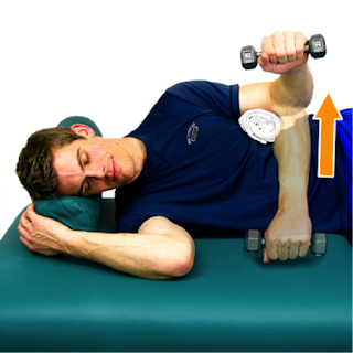
Biceps curl: Stand and hold a 5- to 8-pound weight in your hand. If you do not have a weight, use a soup can or hammer. Bend your elbow and bring your hand (palm up) toward your shoulder. Hold 5 seconds. Slowly straighten your arm and return to your starting position. Do 2 sets of 8 to 12.

Single-arm shoulder flexion:

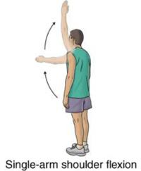
Stand with your injured arm hanging down at your side. Keeping your arm straight, bring your arm forward and up toward the ceiling. Hold this position for 5 seconds. Do 2 sets of 8 to 12. As this exercise becomes easier, add a weight.
Resisted shoulder internal rotation:
Stand sideways next to a door with your injured arm closest to the door. Tie a knot in the end of the tubing and shut the knot in the door at waist level. Hold the other end of the tubing with the hand of your injured arm. Bend the elbow of your injured arm 90 degrees. Keeping your elbow in at your side, rotate your forearm across your body and then slowly back to the starting position. Make sure you keep your forearm parallel to the floor. Do 2 sets of 8 to 12.
Resisted shoulder external rotation:
Stand sideways next to a door with your injured arm farther from the door. Tie a knot in the end of the tubing and shut the knot in the door at waist level. Hold the other end of the tubing with the hand of your injured arm. Rest the hand of your injured arm across your stomach. Keeping your elbow in at your side, rotate your arm outward and away from your waist. Slowly return your arm to the starting position. Make sure you keep your elbow bent 90 degrees and your forearm parallel to the floor. Repeat 10 times. Build up to 2 sets of 15.
Side-lying external rotation:
Lie on your uninjured side with your injured arm at your side and your elbow bent 90 degrees. Keeping your elbow against your side, raise your forearm toward the ceiling and hold for 2 seconds. Slowly lower your arm. Do 2 sets of 15. You can start doing this exercise holding a soup can or light weight and gradually increase the weight as long as there is no pain.


Sleeper stretch:

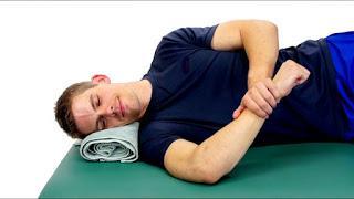
Lie on your injured side with your hips and knees flexed and your arm straight out in front of you. Bend the elbow on your injured side to a right angle so that your fingers are pointing toward the ceiling. Then use your other hand to gently push your arm down toward the floor. Keep your shoulder blades lightly squeezed together as you do this exercise. Hold the stretch for 30 seconds. Repeat 3 times.

Friday, 14 December 2018

Physiotherapy Rehabilitation for Tennis Elbow


               Physiotherapy Rehabilitation for Tennis Elbow

What is Tennis elbow?

Tennis elbow is a condition caused by inflammation of the tendons that attach the extensor carpi radialis brevis to the outer bony prominence (lateral epicondyle) of the elbow. Certain repetitive movements of the wrist can cause this condition. Tennis elbow can occur in anyone who strains the tendons of the forearm and is not limited to tennis players. Tennis elbow is also called lateral epicondylitis.

Why tennis players getting tennis elbow?

Any activity, including playing tennis, that involves repetitive use of the extensor muscles of the forearm can cause acute or chronic tendinitis of the tendinous insertion of these muscles at the lateral epicondyle of the elbow, while attempting One-handed backhand with poor form or technique

Other game players can get tennis elbow?
Yes, You know Sachin Tendulkar, a famous cricketer from India. In 2004, it was happened to him. It was actually a small tear in the origin of muscles at the elbow that runs through to the wrist. In effect, wrist movement caused pain at the elbow. It could happen acutely and immediately, need not have to be over a period of time."

Tennis players what can do to avoid tennis elbow

Hold your injured arm out straight, palm down.Use your other hand to hold the back of your injured arm's hand.Press down so your fingers point to the ground.You should feel a light stretch on the top of your forearm. Do not stretch to the point of pain.

Symptoms:

tenderness over the outside of elbow.
Morning stiffness of elbow or persistent aching
Soreness of Forearm muscles
Elbow pain is severe, while holding or grasping the hand.

Call your health care professional or physiotherapist, if the following conditions develop:
  • Pain that limits your daily activity
  • Pain that lasts despite ice, resting, and over-the-counter anti-inflammatory pain relievers
  • Any weakness or numbness in the hand, which may mean you have another type of injury in the wrist or elbow

Are There Home Remedies for Tennis Elbow?

  • Home remedies include icing the area for 20 minutes twice a day to help to decrease inflammation and relieve pain. Freezing water in a paper cup and tearing away the top rim as the ice melts is an easy way to use ice. Do not put ice directly on the skin. Wrap it in a towel.
  • Rest the sore area to prevent further injury and decrease pain.
  • Over-the-counter anti-inflammatory medications such as ibuprofen, asprin or naproxen may help decrease the pain and swelling and help the healing. 
Physiotherapy intervention:

The first steps in treating tennis elbow are reducing inflammation and resting the irritated muscles and tendons. Ice and compression may also help reduce inflammation and pain. Once inflammation subsides, you can begin gentle exercises to strengthen the muscles of the forearm and prevent recurrence.

Most cases of tennis elbow respond to rest, ice, rehab exercises, pain medicine, and counterforce braces. This injury does take from 6 months to 12 months to heal. Patience helps. Surgery is considered as a last resort when all other nonsurgical treatments have failed.


Treatment for Tennis Elbow:
  • Icing the elbow to reduce pain and swelling. Experts recommend doing it for 20 to 30 minutes every 3 to 4 hours for 2 to 3 days or until the pain is gone.
  • Using an elbow strap to protect the injured tendon from further strain.
  • Taking nonsteroidal anti-inflammatory (NSAID's), such as ibuprofen, naproxen, or aspirin, to help with pain and swelling. However, these drugs can cause side effects, such as bleeding and ulcers. You should only use them occasionally, unless your doctor says otherwise, since they may delay healing.
  • Performing range of motion exercises to reduce stiffness and increase flexibility. Your doctor may recommend that you do them three to five times a day.
  • Getting physiotherapy to strengthen and stretch the muscles. 
  • Strengthening exercises for Tennis Elbow
  •     Ball or sock squeeze


     Hold a tennis ball (or a rolled-up sock) in your hand.

  • Make a fist around the ball (or sock) and squeeze.
  • Hold for about 6 seconds, then relax for up to 10 seconds.
  • Repeat 8 to 12 times.
  • Switch the ball (or sock) to your other hand and do 8 to 12 times.
  • Wrist deviation
    1. Sit so that your arm is supported but your hand hangs off the edge of a flat surface, such as a table.
    2. Hold your hand out like you are shaking hands with someone.
    3. Move your hand up and down.
    4. Repeat this motion 8 to 12 times.
    5. Switch arms.
    6. Try to do this exercise twice with each hand.
    Wrist curls
    1. Place your forearm on a table with your hand hanging over the edge of the table, palm up.
    2. Place a 1- to 2-pound weight in your hand. This may be a dumbbell, a can of food, or a filled water bottle.
    3. Slowly raise and lower the weight while keeping your forearm on the table and palm facing up.
    4. Repeat this motion 8 to 12 times.
    5. Switch arms, and do steps 1 through 4.
    6. Repeat with your hand facing down toward the floor. Switch arms.
    Biceps curls

    1. Sit leaning forward with your legs slightly spread and your left hand on your left thigh.
    2. Place your right elbow on your right thigh, and hold the weight with your forearm horizontal.
    3. Slowly curl the weight up and toward your chest.
    4. Repeat this motion 8 to 12 times.
    5. Switch arms, and do steps 1 through 4.

    Wednesday, 12 December 2018

    Why Athletes with back pain is not getting better?


               Why Athletes with back pain is not getting better?

    All of us treat back pain in our practices.  It can easily be as much as 50% of our patient population, which has led to many schools of thought.  Do I manipulate?  Do I use the algorithms from McKenzie?  Should I strengthen the core?  All of these have their time and place and are each incredibly valuable for the right patient!
    For years, I used these schools of thoughts, but still often struggled with a specific population – RUNNERS. Runners are supposed to come in with aching knees, IT Band Syndrome, Achilles Tendon pain, but back pain?  Yes, more often than you might think, they complain of back pain.  It’s not that manipulating a runner’s back is not helpful and plenty of us could benefit from a few extra planks, but there’s something more.  If you don’t address this, the likelihood of the back pain returning is high because you haven’t addressed the activity provoking the pain.
    Many runners adopt a hyper lordotic position when they are running and even worse, they hinge in a specific segment of their lumbar spine.   They run very upright and may even show excessive vertical oscillation (bounce when they run).
    Once you determine that this may be a contributing factor, you need to look at what may be the cause. Typically, it is one of 3 things:
    1. Habit: The runner is just too upright and needs to work on a forward lean.  You can cue the the runner to fall through their ankles when they run.  Let them stand still with their feet together and begin to fall forward at their ankles.  They then naturally step forward to keep from actually falling. This is will promote the natural forward lean we are looking for.
    2. Hip PROM: Inadequate extension PROM is present, resulting in a compensatory anterior pelvic tilt and hyper lordosis during late stance.
    3. Pelvic and Hip Motor Control: Instead of extending the hips through the gluteals, extension occurs through the lumbar spine and no hip extension PROM deficit is present.
    You can quickly determine whether this is a problem with PROM or motor control by performing both active and passive hip extension in prone.  I often find a PROM restriction exists, but not always.  Once you treat the passive restriction you need to retrain the new motion with motor control drills.  A simple prone hip extension exercise or quadruped hip extension will often do the trick, but you must focus on promoting hip extension and not anterior pelvic tilting.  Finally, retraining running form to use the new ROM and motor control completes the cycle.  You can use your falling drill at this point.

    Monday, 10 December 2018

    New Neuron Generation(Neurogenesis) in Adults

    New Neuron Generation(Neurogenesis) in Adults

    What is adult Neurogenesis?


    "Adult neurogenesis" refers to the ability of the central nervous system (brain and spinal cord) to generate new neurons in adulthood called adult generated neurons. This is as differentiated from "neurogenesis" typically used to describe the processes of neuronal generation that occur during the prenatal (embryonic and fetal) period extending in to the early years of postnatal life.
    Back in the 1800s and the first half of the 20th century, scientists and scholars believed that development of new neurons only occurred early in life and would cease at some point during development, so that no new neurons could be formed in the brain or spinal cord after this 'critical age'.  Some scientists back then disputed this as fact but had no means of disproving the widely accepted notion, the non-availability of advanced equipment back then and less advanced scientific investigation and laboratory techniques meant this notion would go unchallenged for a very long time.

    Definition:
    Adult neurogenesis is the process of generating new neurons which integrate into existing circuits after fetal and early postnatal development has ceased. In most mammalian species, adult neurogenesis only appears to occur in the olfactory bulb and the hippocampus

    What happen in neurological diseases?

    Immense interest has been generated around this area, now that adult neurogenesis is known to be a fact, focus has shifted towards determining the factors that affect adult neurogenesis (increase or decrease it rate of occurrence) and the functions of these adult generated neurons. Interestingly it has been shown that such factors as physical exercise, living in an environmentally enriched area and mentally challenging tasks among other things improve the rate of production of new neurons in the adult hippocampus as well as increase the longevity of these newly generated neurons. In the same vein, scientists have managed to demonstrate that laboratory animals in which the rates of neurogenesis had been increased by such means as physical exercise performed better at tasks such as learning a new skill or spatial navigation in a novel (new) environment. Incidentally, elevated levels of adult hippocampal neurogenesis have been observed in adults with neurological insults and diseases such as stroke (CVA), Alzheimer's disease, Parkinson's disease and dementia among others. These diseases are characterized by death and destruction of neurons and the elevated levels of neurogenesis are hypothesized to be the body's own way of trying to replace the neurons that would have been lost.

    Physiotherapist role in Adult neurogenesis:


    Considering that physical exercise and environmental enrichment will improve the levels of adult neurogenesis, physiotherapists can augment the body's own regenerative capacities by working with patients with neurological diseases to promote activities that enhance hippocampal neurogenesis.

    1. Promote a healthy lifestyle - eat healthy, avoid the use of drugs. The use of drugs such as methamphetamine(reduces the rate of neurogenesis) and decreases cognitive functions
    2. Develop an exercise regime as part of the treatment program for neuro patients.

    So , physiotherapy helps us  in a lot of ways to rehabilitate neurological patients in all ways( also in neurogenesis). Thank you


    Saturday, 8 December 2018

    Rheumatoid Arthritis

                             
                              Rheumatoid Arthritis
    What is Rheumatoid arthritis?
    Rheumatoid arthritis is a chronic inflammatory disorder that can affect more than just your joints. In some people, the condition also can damage a wide variety of body systems, including the skin, eyes, lungs, heart and blood vessels.


    An autoimmune disorder, rheumatoid arthritis occurs when your immune system mistakenly attacks your own body's tissues. Means your immune system attacking your own body tissues. This leads to collapse the joint structure mainly small joints then affecting the major joints like hip and knee sometimes leads to back pain also.


    Causes for RA:


    Rheumatoid arthritis occurs when your immune system     attacks the synovium — the lining of the membranes that surround your joints.In this stage joint space will be widens. The resulting inflammation thickens the synovium, which can eventually destroy the cartilage and bone within the joint. In this stage, joint space will be likened less. In third stage, there is no space in between bones. The tendons and ligaments that hold the joint together weaken and stretch. Gradually, the joint loses its shape and alignment.


    In fact we don't know the exact cause, although a genetic component appears likely. While your genes don't actually cause rheumatoid arthritis, they can make you more susceptible to environmental factors — such as infection with certain viruses and bacteria — that may trigger the disease.


    Risk factors

    Factors that may increase your risk of rheumatoid arthritis include:
    Your sex. Women are more likely than men to develop rheumatoid arthritis. Likely more than 70 percentage.
    Age. Rheumatoid arthritis can occur at any age, but it most commonly begins between the ages of 40 and 60.
    Family history. If a member of your family has rheumatoid arthritis, you may have an increased risk of the disease.
    Smoking. Cigarette smoking increases your risk of developing rheumatoid arthritis, particularly if you have a genetic predisposition for developing the disease. Smoking also appears to be associated with greater disease severity. But actual reason we don't know.
    Environmental exposures. Emergency workers exposed to dust from the collapse of the World Trade Center are at higher risk of autoimmune diseases such as rheumatoid arthritis.Although uncertain and poorly understood, some exposures such as asbestos or silica may increase the risk for developing rheumatoid arthritis.
    Obesity. People who are overweight or obese appear to be at somewhat higher risk of developing rheumatoid arthritis, especially in women diagnosed with the disease when they were 55 or younger.    
    Symptoms

    • Signs and symptoms of rheumatoid arthritis may include:
      • Tender, warm, swollen joints
      • Joint stiffness that is usually worse in the mornings and after inactivity
      • Fatigue, fever and weight loss
    • Early rheumatoid arthritis tends to affect your smaller joints and distal joints first — particularly the joints that attach your fingers to your hands and your toes to your feet.As the disease progresses, symptoms often spread to the wrists, knees, ankles, elbows, hips and shoulders. In most cases, symptoms occur in the same joints on both sides of your body. About 40 percent of the people who have rheumatoid arthritis also experience signs and symptoms that don't involve the joints. Rheumatoid arthritis can affect many non joint structures, including:
      • Skin
      • Eyes
      • Lungs
      • Heart
      • Kidneys
      • Salivary glands
      • Nerve tissue
      • Bone marrow
      • Blood vessels
    • Rheumatoid arthritis signs and symptoms may vary in severity and may even come and go. Periods of increased disease activity, called flares, alternate with periods of relative remission — when the swelling and pain fade or disappear. Over time, rheumatoid arthritis can cause joints to deform and shift out of place.
    • Complications

    • Rheumatoid arthritis increases your risk of developing:
    • Carpal tunnel syndrome. If rheumatoid arthritis affects your wrists, the inflammation can compress the nerve that serves most of your hand and fingers. Who are all having Rheumatoid Arthritis, there is a lot of chances to have a carpel tunnel syndrome.
    • Lymphoma. Rheumatoid arthritis increases the risk of lymphoma, a group of blood cancers that develop in the lymph system , because of auto immune attack.
    • Lung disease. People with rheumatoid arthritis have an increased risk of inflammation and scarring of the lung tissues, which can lead to progressive shortness of breath.
    • Heart problems. Rheumatoid arthritis can increase your risk of hardened and blocked arteries, as well as inflammation of the sac that encloses your heart.
    • Abnormal body composition. The proportion of fat compared to lean mass is often higher in people who have rheumatoid arthritis, even in people who have a normal body mass index (BMI).
    • Infections. The disease itself and many of the medications used to combat rheumatoid arthritis can impair the immune system, leading to increased infections.
    • Dry eyes and mouth. People who have rheumatoid arthritis are much more likely to experience Sjogren's syndrome, a disorder that decreases the amount of moisture in your eyes and mouth.
    • Rheumatoid nodules. These firm bumps of tissue most commonly form around pressure points, such as the elbows. However, these nodules can form anywhere in the body, including the lungs.

    • Osteoporosis. Rheumatoid arthritis itself, along with some medications used for treating rheumatoid arthritis, can increase your risk of osteoporosis — a condition that weakens your bones and makes them more prone to fracture.
    • Diagnosis

    • Rheumatoid arthritis can be difficult to diagnose in its early stages because the early signs and symptoms mimic those of many other diseases. There is no one blood test or physical finding to confirm the diagnosis.
    • Blood tests

    • People with rheumatoid arthritis often have an elevated erythrocyte sedimentation rate (ESR, or sed rate) or C-reactive protein (CRP), which may indicate the presence of an inflammatory process in the body. Other common blood tests look for rheumatoid factor and anti-cyclic citrullinated peptide (anti-CCP) antibodies.
    • Imaging tests

    • Your doctor may recommend X-rays to help track the progression of rheumatoid arthritis in your joints over time. MRI and ultrasound tests can help your doctor judge the severity of the disease in your body.
    • Medications

    • The types of medications recommended by your doctor will depend on the severity of your symptoms and how long you've had rheumatoid arthritis.
      • NSAIDs. Nonsteroidal anti-inflammatory drugs (NSAIDs) can relieve pain and reduce inflammation. Over-the-counter NSAIDs include ibuprofen (Advil, Motrin IB) and naproxen sodium (Aleve).
      • Steroids. Corticosteroid medications, such as prednisone, reduce inflammation and pain and slow joint damage. .
      • Disease-modifying antirheumatic drugs (DMARDs). These drugs can slow the progression of rheumatoid arthritis and save the joints and other tissues from permanent damage.
      • Biologic agents.  this newer class of DMARDs includes abatacept (Orencia), adalimumab (Humira), anakinra (Kineret), certolizumab (Cimzia), etanercept (Enbrel), golimumab (Simponi), infliximab (Remicade), rituximab (Rituxan), tocilizumab (Actemra) and tofacitinib (Xeljanz).
      • Rheumatoid arthritis surgery may involve one or more of the following procedures:
        • Synovectomy. Surgery to remove the inflamed synovium (lining of the joint). Synovectomy can be performed on knees, elbows, wrists, fingers and hips.
        • Tendon repair. Inflammation and joint damage may cause tendons around your joint to loosen or rupture. Your surgeon may be able to repair the tendons around your joint.
        • Joint fusion. Surgically fusing a joint may be recommended to stabilize or realign a joint and for pain relief when a joint replacement isn't an option.
    Total joint replacement. During joint replacement surgery, your surgeon removes the damaged parts of your joint and inserts a prosthesis made of metal and plastic                                                                                                                                                                                                                          A structured exercise program can be greatly beneficial to the overall well-being and functioning of the individual with rheumatoid arthritis. Such a program should focus on stretching, strengthening and aerobic conditioning while conserving energy.