Friday, 11 January 2019

Physiotherapy Management of Shoulder pain in patients with stroke

Management of shoulder pain in patients with stroke:


Shoulder pain affects from 16% to 72% of patients after a cerebrovascular accident. Hemiplegic shoulder pain causes considerable distress and reduced activity and can markedly hinder rehabilitation. The aetiology of hemiplegic shoulder pain is probably multifactorial. The ideal management of hemiplegic stroke pain is prevention. For prophylaxis to be effective, it must begin immediately after the stroke. Awareness of potential injuries to the shoulder joint reduces the frequency of shoulder pain after stroke. The multidisciplinary team, patients, and carers should be provided with instructions on how to avoid injuries to the affected limb. Foam supports or shoulder strapping may be used to prevent shoulder pain. Overarm slings should be avoided. Treatment of shoulder pain after stroke should start with simple analgesics. If shoulder pain persists, treatment should include high intensity transcutaneous electrical nerve stimulation or functional electrical stimulation. Intra-articular steroid injections may be used in resistant cases. 

Image result for management of shoulder pain in patients with stroke


Occurence:
Shoulder pain is a common complication after a cerebrovascular accident. From 16% to 72% of stroke patients develop hemiplegic shoulder pain. It may occur in up to 80% of stroke patients who have little or no voluntary movement of the affected upper limb.
Hemiplegic shoulder pain has been shown to affect stroke outcome in a negative way. It interferes with recovery after a stroke: it can cause considerable distress and reduced activity and can markedly hinder rehabilitation. Royet al demonstrated that the presence of hemiplegic shoulder pain is strongly associated with prolonged hospital stay and poor recovery of arm function in the first 12 weeks after stroke.
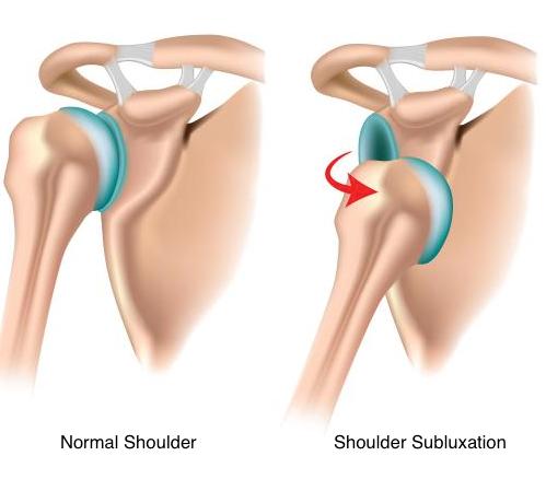
Image result for prevention of shoulder pain in patients with stroke
The cause of hemiplegic shoulder pain is the subject of considerable controversy. The following processes have all been postulated as causes of a painful hemiplegic shoulder: glenohumeral subluxation, spasticity of shoulder muscles, impingement, soft tissue trauma, rotator cuff tears, glenohumeral capsulitis, bicipital tendinitis, and shoulder hand syndrome.Traction neuropathy of the brachial plexus may also play a part. Unusual patterns of motor recovery or spasticity or unusually severe focal atrophy may suggest brachial plexus injury. Poor handling of a hemiplegic limb may exacerbate a pre-existing condition such as osteoarthritis. Thus, pre-morbid disease of the shoulder may predispose to hemiplegic shoulder pain. Stroke patients may suffer from pain that is caused by the stroke itself (central post-stroke pain). The role of central post-stroke pain in the aetiology of hemiplegic shoulder pain is uncertain. Abnormal tone (both spasticity and flaccidity) has been suggested as an aetiological factor in hemiplegic shoulder pain. However, clinical observations suggest that shoulder pain does not occur until spasticity develops. Most authorities agree that the aetiology of hemiplegic shoulder pain is probably multifactorial.

Prevention:

The ideal management of hemiplegic stroke pain is to prevent it happening in the first place. Various strategies have been employed in the prophylaxis of hemiplegic shoulder pain. For prophylaxis to be effective, it must be begin immediately after the stroke. Once the patient has pain, resultant anxiety and overprotection will follow.

HANDLING

Poor handling and positioning of the affected upper limb in stroke patients contribute toward shoulder pain.
The mobility of the recovering stroke patient is dependent on the assistance of nurses, therapists, doctors, other ancillary staff, and family members. It is also dependent on his/her own efforts. Handling, positioning, and transferring on a day-to-day basis can exert great stress on the vulnerable shoulder. The problem may be exacerbated by the patient's sensory and perceptual deficits. There has been concern that trauma to the constituent components of the shoulder joint may be caused by poor handling of the patient's affected arm.
Wanklyn et al studied the prevalence of hemiplegic shoulder pain and associated factors in patients with stroke. Sixty three per cent of the patients developed hemiplegic shoulder pain in the first six months after their stroke. Patients who needed help with transfers were more likely to develop hemiplegic shoulder pain. Certainly, patients with markedly decreased voluntary movement after a cerebrovascular accident frequently experience shoulder joint malalignment or subluxation in the early stages of recovery.
Careful positioning and handling of the limb are thought to prevent hemiplegic shoulder pain, but there is a range of opinions about how correct limb positioning is best achieved.
Braus et al investigated the efficacy of an information and education programme in the prevention of hemiplegic shoulder pain. All members of the diagnostic and therapeutic team as well as patients and their family were provided with instructions on how to avoid injuries to the affected limb. The investigators found that awareness of potential injuries to the structures of the shoulder joint reduced the frequency of shoulder pain from 27% to 8%.
Fitzgerald-Finch et al advocated the use of the Australian lift when handling these patients: they felt it to be of value as the weight of the patient is taken on the shoulders of the carer and the patient's shoulder is protected.

STRAPPING

Glenohumeral joint subluxation may be a contributing factor in the development of shoulder pain in this group of patients. Shai et alhypothesised that earlier radiological diagnosis of subluxation might enable more effective prevention than if it is delayed. However, this has not been proved. Despite this, a variety of slings have been designed to try to correct subluxation and pain in stroke patients with hemiplegia. Not all such devices have been successful: supportive devices developed by Buccholtz Moodie et al and Williams et al were not proved to be effective in correcting subluxation of the shoulder.Image result for prevention of shoulder pain in patients with stroke
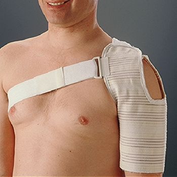
Physiotherapists have employed various forms of strapping designed for shoulder pain or subluxation after a cerebrovascular accident. Unfortunately, the effectiveness of many of these strapping methods remains largely unproved. Ancliffe undertook a pilot study to determine the effectiveness of a strapping technique to prevent shoulder pain after a stroke. The pilot study demonstrated that strapping the hemiplegic shoulder delayed the onset of shoulder pain. In patients with subluxation and shoulder pain, use of a Varney brace has been reported to be successful: patients become asymptomatic within seven days.
Image result for strapping of shoulder pain in patients with stroke
External support can be discontinued when muscle tone around the glenohumeral joint is sufficient to prevent subluxation. An exercise programme should always accompany the use of a sling.
However, a number of authors have reported that slings may hold the limb in a poor position that is likely to cause soft tissue contracture and have an adverse effect on symmetry, balance, and body image.

PHYSIOTHERAPY

Some studies have noted that passive abduction of the hemiplegic arm can result in rotator cuff injury: this in turn causes shoulder pain.
However, therapeutic range of motion exercises done by the patients can involve passive abduction of the arm. Kumar et alanalysed the occurrence of pain in patients receiving three different rehabilitation exercise programs: range of motion by the therapist, use of a skateboard, and use of an overhead pulley. They found that patients who used the overhead pulley had the highest risk of developing shoulder pain and concluded that use of the pulley should be avoided during stroke rehabilitation.
Image result for physiotherapy for shoulder pain in patients with stroke
If impingement during range of motion exercises is determined to be the cause of hemiplegic shoulder pain, the amplitude of passive movement should be kept within the pain-free range. Caldwellet al reported that pain subsided in 43% of patients with hemiplegic shoulder pain when the amplitude of passive range of motion was reduced.
Increase in the prevalence of shoulder pain in the first weeks after discharge in patients who did not continue to exercise properly.

Treatment

Radiological investigations should exclude dislocation or fracture of the shoulder before further management is instigated. Various treatments have been suggested as being beneficial in shoulder pain after stroke: these include physiotherapy, localised cooling, infrared, ultrasound, and intra-articular injections of steroids and local anaesthetics. Until recently there has been a shortage of prospective controlled clinical trials.

PHYSIOTHERAPY

Physiotherapy has been used in the treatment of hemiplegic shoulder pain. There are two major approaches to therapy in this field: those that focus on the problem as a localised mechanical one; and those that view the problem as a neurological one. Local treatments used have included heat and cold therapy. Slings and shoulder supports have also been used. Positioning is also considered important by many authors. Other physiotherapy approaches include those of Bobath, Brunnstrom, and proprioceptive neuromuscular facilitation. Until recently, the evidence for the effectiveness of these methods of physiotherapy has been poor. Partridge examined the effectiveness of two methods of physiotherapy in the treatment of hemiplegic shoulder pain: cryotherapy or the Bobath approach. The cryotherapy approach involved the application of ice to the affected shoulder. The Bobath approach is a neurologically based holistic approach that is frequently used in the UK.
There were no significant differences between the two treatments in terms of severity of pain at rest or on movement or for reported distress. However the proportion of patients who reported no pain after treatment was greater in those who received the Bobath approach.

TRANSCUTANEOUS ELECTRICAL NERVE STIMULATION


Leandri et al evaluated the effectiveness of high intensity versus low intensity transcutaneous electrical nerve stimulation (TENS) versus placebo for patients with hemiplegic shoulder pain. Low intensity TENS involves electrical stimulation just above the level of the skin sensory threshold. High intensity TENS is sufficient to elicit muscle contraction and an almost painful sensation. The investigators found that patients who received high intensity TENS had significant improvements in passive range of motion for flexion, extension, abduction, and external rotation at the shoulder. The patients who received high intensity TENS also reported very satisfactory pain relief.

FUNCTIONAL ELECTRICAL STIMULATION

There are have been a number of studies of the effectiveness of functional electrical stimulation (FES) in shoulder pain after stroke.
Faghri et al studied the effects of a FES treatment programme designed to prevent glenohumeral joint stretching and subsequent subluxation and shoulder pain in stroke patients. They demonstrated a beneficial effect on subluxation and improvement in other parameters such as pain, range of motion, and arm function.
Chantraine et al completed a long term controlled study of the use of FES in hemiplegic stroke patients diagnosed with a subluxed and painful shoulder. They found that a 24 month FES programme was effective in reducing the severity of subluxation and pain and may have facilitated recovery of shoulder function in these patients.
As an extremely mobile joint, the shoulder sacrifices stability for mobility.  Basmajian determined through electromyographic studies that the supraspinatus, and to a lesser extent the posterior deltoid muscles, played a key part in maintaining glenohumeral alignment. Chaco and Wolf also demonstrated the importance of the supraspinatus muscle in preventing downward subluxation of the humerus. Two studies have investigated the application of electrical stimulation to the supraspinatus and posterior deltoid muscles: Baker and Parker demonstrated the beneficial effects of FES in stroke patients with a chronic shoulder dislocation. However, patients deteriorated after withdrawal from treatment though not back to pre-treatment levels. Linn et al carried out a prospective randomised study to determine the efficacy of FES in the prevention of shoulder subluxation in stroke patients. They found that FES does prevent shoulder subluxation, but this effect was not maintained after the withdrawal of treatment.

Wednesday, 9 January 2019

Positioning of Stroke patients

Introduction:

The aim of positioning the patient is to try to promote optimal recovery by modulating muscle tone, providing appropriate sensory information, increasing spatial awareness and prevention of complications such as pressure sores, contracture, pain, respiratory problems and assist safer eating.
Correct positioning can help to reduce the risk of;
AspirationContracture Pressure AreasShoulder Pain Swelling of the Extremities 

Image result for positioning for stroke patients in bed

Aims of positioning:

Normalise Tone or Decrease Abnormal influence on the Body
Maintain Skeletal Alignment
Prevent, Accommodate or Correct Skeletal Deformity
Provide Stable Base of Support
Promote Increased Tolerance of Desired Position
Increased Stimulation to Affected Side
Increased Spatial Awareness
Promote Patient Comfort
Facilitate Normal Movement Patterns
Control Abnormal Movement Patterns
Manage Pressure
Decrease Fatigue
Enhance Autonomic Nervous System Function (Cardiac, Digestive and Respiratory Runction)
Facilitate Maximum Function
Improved Ability to Interact with the Environment

Types of positioning:

1Sitting in a Chair or Sitting in a Wheelchair:Image result for Sitting in a Chair or Sitting in a Wheelchair

It is vital that as soon as the person is capable of sitting out that they are facilitated to do so. Sitting out is essential to build up tolerance; provide maximum stimulation; give a sense of normality.

Head over PelvisHips at 90 degreesKnees at 90 degreesSlight extension of lumber regionFeet in neutral position and supportedWeight evenly distributed between both buttocksArm should be protracted forward and supported

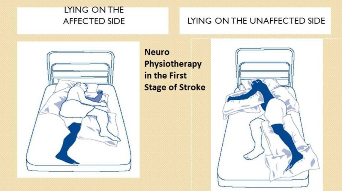
2. Side lying on the unaffected side:

The stroke arm should be well forward, keeping the elbow straight and supported on a pillow.
The stroke leg should be brought far enough in front of the body to prevent the patient rolling on to the back, the knee bent and leg supported on a pillow.
A small pillow can then be placed under the patient's waist to maintain the the line of the spine.
When lying on the side position, the patient should have two pillows only under the head. 
Image result for Side lying on the unaffected side

3. Side lying on the affected side:

This should always be encouraged with the stroke shoulder well forward so that the body weight is supported on the flat of the shoulder blade and not on the point of the shoulder.
One or two pillows for head
Place the stroke leg with the thigh so that it is in line with the trunk, and bend the knee slightly.
The unaffected leg should be brought forward and placed with the knee bent on a pillow in front of the affected leg for comfort. This prevents the patient rolling onto his back.
Lastly, bend the head forward a little.  

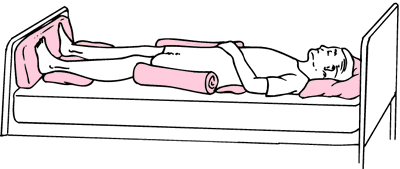
4.Lying supine:

This is the position most likely to encourage spasticity, but some patients do like to lie on their back for a while and it will be required for some treatments.Image result for Lying supine


Place two pillows under the patient's head and help him/her bend their head slightly towards their unaffected shoulder and gently turn their head towards their stroke side but do not uses force.
A small pillow is placed under the buttock of the stroke side and should extend just to the knee, this will relax the leg and prevent it turning out at the hip.
A pillow is placed under the stroke arm which is kept straight at the elbow and if possible, the palms of the hand facing upwards.
The bed must be the correct height to promote independence and safety for the patient, family and health care workers. 

5. Sitting up in bed:

Sitting in bed is desirable for short periods only
Must be upright and well supported with pillows
Consider extra support using pillows under arms or knees

Sitting versus lying:

When seated, nearly half of the body weight is supported on 8% of the sitting areas at or near the ischial tuberosities.
Therefore, interface pressures are much higher in sitting than lying

Distribution of weight when seated normally:

Buttocks & Thighs 75%
Feet flat on floor 19%
Back 4%
Arms 2%
Total 100%

Pressure relief:


 A person who has had a stroke may be susceptible to developing pressure sores 
Assess the person and decide on an appropriate cushion use in order to
1.Prevent further skin breakdown,
2.To assist with healing
3.To facilitate the patient to sit out as much as possible

Monday, 7 January 2019

Effectiveness of Meckenzie Approach in Back Pain

The McKenzie assessment consists of taking a patient history and performing a physical exam. Both are used to gauge the degree of impairment as well as identify any red flags that might be contrary to exercise-based treatment (e.g. fracture, tumor, infections, or systemic inflammatory disease).
During the McKenzie physical examination, patients are taken through provocative loading strategies (movements) that help classify the patient and determine the best treatment approach. The movements are intended to either increase or decrease symptoms. For example, patients may be asked to perform single and/or repeated flexion or extension movements forward and backward

Image result for mckenzie approach
The McKenzie method is a classification system and a classification-based treatment for patients with low back pain. A acronym for the McKenzie method is mechanical diagnosis and therapy (MTD). The McKenzie method was developed in 1981 by Robin McKenzie, a physical therapist from New Zealand.
The McKenzie method exists of 3 steps: evaluation, treatment and prevention. The evaluation is received using repeated movements and sustained positions. With the aim to elicit a pattern of pain responses, called centralization, the symptoms of the lower limbs and lower back are classified into 3 subgroups: derangement syndrome, dysfunction syndrome and postural syndrome. The choice of exercises in the McKenzie method is based upon the direction (flexion, extension or lateral shift of the spine). The aims of the therapy are: reducing pain, centralization of symptoms (symptoms migrating into the middle line of the body) and the complete recovery of pain. The prevention step consists of educating and encouraging the patient to exercise regularly and self-care. All exercises for the lumbar spine are repeated a number of times to end-range on spinal symptoms in one direction. When you do only 1 repetition, this will cause pain. When you repeat it several times the pain will decrease. Also after movement termination the changes in pain intensity can persist, which leads to a treatment modality. A single direction of repeated movements or sustained postures leads to sequential and lasting abolition of all distal referred symptoms and subsequent abolition of any remaining spinal pain.

Classification:

McKenzie described in 1981 for the first time the mechanical classification in the McKenzie system. The patients are classified into four groups according to the mechanical and symptomatic response to repeated movements and sustained positions. Each syndrome demands a different management approach. In the paragraph below you will find the four categories of the McKenzie classification with their descriptions

Posture syndrome:

Refers to pain which occurs due to a mechanical deformation of normal soft tissue from prolonged end range loading of periarticular structures.

The pain arises during static positioning of the spine: for example sustained slouched sitting.

The pain disappears when the patient is moved out of the static position.

The treatment includes: patient education, correction of the posture by improving posture by restoring lumbar lordosis, avoiding provocative postures and avoid prolonged tensile stress on normal structure.

Dysfunction syndrome:

  • Refers to pain which is a result of mechanical deformation of structurally impaired tissues like scar tissue or adhered or adaptively shortened tissue.
  • The pain arises at the end range of a restricted movement.
  • The treatment includes:
Mobilizing exercises in the direction of the dysfunction or in the direction that reproduces the pain. The aim is to remodel that tissue, which limits the movement, through exercises so that it becomes pain-free over 
Derangement syndrome;
Is the most prevalent treatment classification. Refers to pain which is caused by a disturbance in the normal resting position of the affected joint surfaces.This syndrome is classified in two groups.

1. Irreducible derangement:

The criteria for derangement are present.No strategy is capable to produce a permanent change in symptoms.

2. Reducible derangement:

Shows one direction of repeated movement which decreases or centralizes referred symptoms = preferred direction.

Shows also an opposite repeated movement characterized by production or increase or distal movement of the symptoms.

The treatment includes: examination of the patient’s symptomatic and mechanical response to repeated movements or sustained positions because the chosen treatment depends on the clinically induced directional preference.

Others:

Contains minority of patients who do not fit within one of the three mechanical syndromes but who demonstrate symptoms and signs of other pathology like:
Spinal stenosis
Hip disorders
Sacroiliac disorders
Low back pain in pregnancy
Zygapophyseal disorders
Spondylolysis and spondylolisthesis
Post-surgical problems. 

Management:

Unlike other exercises for treating low back pain meant for muscle strengthening, stability and restoring range of motion, the McKenzie method exercises are meant to directly diminish or even eliminate the patients symptoms. This effect is accomplished by providing corrective mechanical directional movements in end range. The McKenzie method educates patients regarding movement and position strategies can reduce pain. A cautious progression of repeated forces and loads is used in this method.  The exercises may be uncomfortable at first, but after some repetitions the symptoms will decrease. 
  • Principles:
    • Kyphotic antalgic management: extension principle
    • Acute coronal antalgic management: lateral flexion-then-extension principle
    • Acute lordotic antalgic management: Flexion principle.
Example:

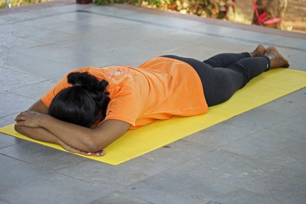
1. Lying prone:

The patient takes place at the treatment table in prone position. The arms have to be parallel with the thorax, with the hands next to the pelvis. The head is turned to one side. This position creates automatically a lordosis of the lumbar spine. Patients with posterior derangement should be careful when arising from the position to standing. It is important that, while arising, the restored lordosis is maintained. In any kind of derangement it is important to perform the exercise long enough (5-10 minutes) for the fluid to alter its position anteriorly. In minor derangement, prone lying may reduce the derangement without any other procedures being required.

Image result for lying prone Although this position may be painful, the pain does not indicate the procedure is undesirable if it is felt centrally. In major derangement, for example patients with lumbar kyfosis, it is possible that the patients cannot tolerate the prone position unless they are lying over a few pillows. In case of dysfunction the loss of extension may be enough to prevent lying prone because the soft tissue shortening has reduced the range of motion and extension stress produces pain.

2. Extension in lying:


The patient lies on his abdomen while the hands are placed near the shoulders. The hands are placed with the palms down. Now the patient makes a press-up movement with straight arms. The Pelvis stays near the table while the patient presses the thorax upwards. After this movement the patient returns to his starting position and repeats this exercise 10 times. The first couple of exercises have to be done easily, but after a few times the movement has to be made to the maximum extension range that is possible. The aim of this exercise is to make the lumbar spine relax after the maximum extension, in the relaxation phase. The maximum degree of extension is obtained with this exercise. It is possible that there occurs central low back pain described as a strain pain, but it will gradually wear off. An intermittent extension stress is influencing the contents and surrounding structures of the lumbar segments, having a pumping as well as a stretching effect. This procedure is the most important and effective in the treatment of derangement as well as extension dysfunction.

3. Extension in standing:


The patient stands up straight with his feet apart, to remain a stable position. The hands are placed on the lumbar region, in the area of the spina iliaca posterior superior. His hands fixate the pelvis while the patient leans backwards. The patient has to lean backwards as far as possible. This exercise has to be repeated ten times. It has similar effects on derangement and dysfunction as extension in lying. In derangement, extension in standing is designed to reduce accumulation of nuclear material in the posterior compartment of the intervertebral joint. The procedure is important in the prevention of the onset of low back pain during or after prolonged sitting and is very effective when performed before pain is actually felt.

4. Rotation mobilization in extension:

The patient lies in a prone position on the treatment table with his arms parallel with the trunk and the head turned to one side. The therapist stands next to the patient and places the heels of the hands on the lumbar region. One will fixate the processus transverses of the vertebra on top of the vertebra you want to rotate. The other hand will make a rotation of the vertebra beneath in the opposite direction. This is more a technique than an exercise, but has to be repeated also ten times. In derangement rotation mobilization in extension has to be performed first to bring about centralization of nuclear material in the disc. Followed by symmetrical extension mobilization to restore the nucleus to its more anterior position. In derangement mechanical deformation is extremely undesirable. In dysfunction an increase of deformation with certain limits is desirable.

5. Self Treatment Exercises:

  • Rest position for cold pack
  • Sphinx-movement
  • standing back extension
  • Pelvic side shift
This exercise is called a “mirror exercise” and can be helpful when you have a “blocked” back and you’re leaning to one side because of it. The patient has to lean with his upper body against the wall, while his feet take same distance from the wall. Now the patient has to move his pelvis against the wall and back to the beginning position. This exercise has to be repeated 8-10 times.

Effectiveness:

For acute pain:
  1. The McKenzie Method is not clinically superior to all interventions in treating acute LBP but is more effective at reducing pain intensity when compared to manual therapy and exercise combined.
For chronic pain:
  1. The McKenzie Method was more effective at reducing pain and disability than “other” interventions,
  2. McKenzie Method was more effective at reducing disability but not pain when compared to exercise alone,
  3. McKenzie Method was not more effective than a combined exercise, manual therapy and education intervention.
As always take these findings with a pinch of salt. The effects of the McKenzie Method could be as a result of the fact large groups of patients may not fall into their subgroups and would have benefited from any exercise anyway.  The McKenzie Method is a mechanical-based system and does not account for psychological aspects of pain or alternative theories of pain. It lumps you into a category, disregards psychological aspects or more nuanced aspects of treatments. Still this doesn’t mean McKenzie isn’t a viable option for treatment. Just bare this in mind. Other treatments and assessment tools / strategies have proven as or more effective.

Saturday, 5 January 2019

Myofacial Pain Syndrome

Myofascial pain syndrome is a chronic pain disorder. In this condition, pressure on sensitive points in your muscles (trigger points) causes pain in the muscle and sometimes in seemingly unrelated parts of your body. This is called referred pain.
This syndrome typically occurs after a muscle has been contracted repetitively. This can be caused by repetitive motions used in jobs or hobbies or by stress-related muscle tension.


While nearly everyone has experienced muscle tension pain, the discomfort associated with myofascial pain syndrome persists or worsens. Treatment options include physical therapy and trigger point injections. Pain medications and relaxation techniques also can help.

Symptoms

Signs and symptoms of myofascial pain syndrome may include:
  • Deep, aching pain in a muscle
  • Pain that persists or worsens
  • A tender knot in a muscle
  • Difficulty sleeping due to pain

Causes

Sensitive areas of tight muscle fibers can form in your muscles after injuries or overuse. These sensitive areas are called trigger points. A trigger point in a muscle can cause strain and pain throughout the muscle. When this pain persists and worsens, doctors call it myofascial pain syndrome.

Risk factors

Myofascial pain syndrome is caused by a stimulus, such as muscle tightness, that sets off trigger points in your muscles. Factors that may increase your risk of muscle trigger points include:
  • Muscle injury. An acute muscle injury or continual muscle stress may lead to the development of trigger points. For example, a spot within or near a strained muscle may become a trigger point. Repetitive motions and poor posture also may increase your risk.
  • Stress and anxiety. People who frequently experience stress and anxiety may be more likely to develop trigger points in their muscles. One theory holds that these people may be more likely to clench their muscles, a form of repeated strain that leaves muscles susceptible to trigger points.

Complications

Complications associated with myofascial pain syndrome may include:

Sleep problems. Signs and symptoms of myofascial pain syndrome may make it difficult to sleep at night. You may have trouble finding a comfortable sleep position. And if you move at night, you might hit a trigger point and awaken.

Fibromyalgia. Some research suggests that myofascial pain syndrome may develop into fibromyalgia in some people. Fibromyalgia is a chronic condition that features widespread pain. It's believed that the brains of people with fibromyalgia become more sensitive to pain signals over time. Some doctors believe myofascial pain syndrome may play a role in starting this process.

Diagnosis

During the physical exam, your doctor may apply gentle finger pressure to the painful area, feeling for tense areas. Certain ways of pressing on the trigger point can elicit specific responses. For instance, you may experience a muscle twitch.

Muscle pain has many possible causes. Your doctor may recommend other tests and procedures to rule out other causes of muscle pain.

Treatment

Treatment for myofascial pain syndrome typically includes medications, trigger point injections or physical therapy. No conclusive evidence supports using one therapy over another, but exercise is considered an important component of any treatment program. Discuss your options and treatment preferences with your doctor. You may need to try more than one approach to find pain relief.
  • Medications

    Medications used for myofascial pain syndrome include:
    • Pain relievers. Over-the-counter pain relievers such as ibuprofen (Advil, Motrin IB, others) and naproxen sodium (Aleve) may help some people. Or your doctor may prescribe stronger pain relievers. Some are available in patches that you place on your skin.
    • Antidepressants. Many types of antidepressants can help relieve pain. For some people with myofascial pain syndrome, amitriptyline appears to reduce pain and improve sleep.
    • Sedatives. Clonazepam (Klonopin) helps treat the anxiety and poor sleep that sometimes occur with myofascial pain syndrome. It must be used carefully because it can cause sleepiness and can be habit-forming.

    Therapy

    A physical therapist can devise a plan to help relieve your pain based on your signs and symptoms. Physical therapy to relieve myofascial pain syndrome may involve:
    • Stretching. A physical therapist may lead you through gentle stretching exercises to help ease the pain in your affected muscle. If you feel trigger point pain when stretching, the physical therapist may spray a numbing solution on your skin.
    • Posture training. Improving your posture can help relieve myofascial pain, particularly in your neck. Exercises that strengthen the muscles surrounding your trigger point will help you avoid overworking any one muscle.
    • Massage. A physical therapist may massage your affected muscle to help relieve your pain. The physical therapist may use long hand strokes along your muscle or place pressure on specific areas of your muscle to release tension.
    • Heat. Applying heat, via a hot pack or a hot shower, can help relieve muscle tension and reduce pain.
    • Ultrasound. This type of therapy uses sound waves to increase blood circulation and warmth, which may promote healing in muscles affected by myofascial pain syndrome.

    Needle procedures

    Injecting a numbing agent or a steroid into a trigger point can help relieve pain. In some people, just the act of inserting the needle into the trigger point helps break up the muscle tension. Called dry needling, this technique involves inserting a needle into several places in and around the trigger point. Acupuncture also appears to be helpful for some people who have myofascial pain syndrome

Thursday, 3 January 2019

Impacts of Aerobic Exercises

AEROBIC EXERCISES:

                       Aerobic exercise is sometimes known as "cardio"- exercise that requires pumping of oxygenated blood by the heart to deliver oxygen to working muscles. Aerobic exercise stimulates the heart rate and breathing rate to increase in a way that can be sustained for the exercise session. Health benefits. Besides strengthening your heart and lungs, aerobic exercise can help lower your cholesterol, reduce your risk of type 2 diabetes, improve your immune function, and lower your blood pressure. Physical benefitsAerobic exercise burns up calories, which can in turn help you shed excess weight.


Benefits of Aerobics:

                 Regular aerobic exercise has significant cardiovascular benefits, including a reduction in incidence of and mortality from coronary artery disease--probably because of positive effects on blood lipid levels and blood pressure. Aerobic exercise can also be an important adjunct to a weight-loss program.

                 Aerobic exercises are brisk exercises, such as running or swimming, that make your heart and lungs work hard, increasing the amount of oxygen circulating through your blood. To lose weight with aerobics, you'll need to burn more calories than you consume and exercise regularly.

                 Truly any exercise is going to help you lose weightAerobic exercise will assist in weight loss due to the amount of calories that are burned. You may potentially burn more calories through intense aerobic exercise than what you burn during anaerobic exercise. However, anaerobic exercise will also help in weight loss.

Aerobic exercises are, Walking, Running, Swimming, Aquarobics, Cycling, Rowing, Boxing, Aerobic and Cardio classes etc

Tuesday, 1 January 2019

Erb's Palsy


ERB'S PALSY(BRACHIAL PLEXUS BIRTH INJURY)
                                                    Erb's Palsy or Erb–Duchenne palsy is a paralysis of the arm caused by injury to the upper group of the arm's main nerves, specifically the severing of the upper trunk C5–C6 nerves. These form part of the brachial plexus, comprising the ventral rami of spinal nerves C5–C8 and thoracic nerve T1.

Causes:
                             They are usually due to trauma, for example falling on the shoulder, or traction on the arm at birth - in which case the name Erb Duchenne paralysis is given.
Specified features:
                            The arm hangs at the side with the elbow extended and the forearm pronated; the so-called waiter's tip or Erb's palsy. Like policemen tip getting position, So it's called as "policemen tip palsy"

PHYSICAL THERAPY  REHABILITATION:
                                If muscle having  less than 1 power means , Physios are using some of muscle activation activities are, MASSAGE, QUICK ICE, BRUSHING, ELECTRICAL MUSCLE STIMULATION, QUICK STRETCH etc..

If the muscles are having power more than 1 , we are using some of muscle strengthening  exercises ,
                                
1.Activities and exercises to promote recovery of movement and muscle strength 

2.Exercises to maintain range of movement in the joints to prevent stiffness and pain 

3.Exercises to promote increased awareness of the arm 

4.Provision of splints to prevent secondary complications and improve function