Tuesday, 26 March 2019

W sitting is normal?

W sitting and its impact


W sitting:


W sitting is when a child sits on their bottom with their knees bent and feet positioned outside of their hips. If you're standing above your child, you will see their legs and body make the shape of a W.


Many parents have heard the phrase “W sitting” and that it is “bad” for their child to sit this way. However, many are unaware of the reason that children are discouraged from sitting in this position.


First of all – what is W sitting?


W Sitting is when a child is sitting on their bottom with both knees bent and their legs turned out away from their body. If you were to look at the child from above their head, his or her legs will be in the shape of the letter “W.” Their knees and thighs may be touching together or spread apart.
For many children, this is a preferred or comfortable position, and they sit that way without even thinking about it. Often times, kids who sit in this position are doing so in order to make up for weaknesses they may have in their hips and trunk. The added stability of this position allows them to play with toys in an upright sitting position without worrying about falling over.
                         Reasons for w sitting:
They're just responding to their natural anatomy.” If your child wants to sit in a W position, it means there's no excessive stress on his joints, muscles or knees because kids know how to avoid pain in their bodies, she added. A child cannot dislocate his hip by sitting this way, both doctors said.

Common reasons why children W-sit that SHOULD BE ADDRESSED:

  • Limited core strength: The W-sitting position gives kids a wider base of support. This may be used to compensate for weak belly and back muscles that make it tiring or challenging to sit in other positions.
  • Muscle tightness: Tight muscles of the legs (particularly the hamstrings on the backs of the thighs) and hips can lead a child to prefer a W-sitting position over long sitting (legs stretched out in front) or tailor sitting ("criss cross applesauce" position).
  • Low muscle tone: We often talk about muscle strength, which refers to active, contracted muscles. Muscle tone is the resting state of the muscles and is controlled by the brain. Some kids have what's called hypotonicity, or low tone. When they aren't actively firing their muscles, these kids have floppier, softer muscles that have a harder time holding their bodies upright. W-sitting is very often seen in kiddos with low muscle tone.
  • Poor trunk rotation skills: If a child is lacking the ability to twist the torso adequately, he will struggle to transition into and out of a sitting position on his bottoms and may compensate by W-sitting.

Common reasons why children W-sit that are TOTALLY NORMAL:

  • Fine motor control: Children (and adults) get the most coordinated, controlled movements of the hands and fingers when they are really steady through the body and arms. It is normal that at times your child will assume the most stable sitting position possible when completing challenging fine motor tasks.
  • Flexibility: Let's face it, most adults don't W-sit in part because we can't. Most children have the flexibility to easily assume a W-sit position and so they likely will at times.
  • Convenient transitions: This one applies particularly to babies. The quickest way to find a seat while crawling is to plop the bootie down into a W-sit. Sure, your baby could rotate and get into a nice seated position but when the dog barking grabs her attention mid-crawl, it makes sense that she finds the quickest transition possible to sit up and check things out. Similarly, if your baby plays in a tall kneel position (balancing on the knees, shins and tops of feet with bottom lifted), she'll likely drop down and back to sit on her calves and possibly to W-sit. It's a natural, easy transition.
It is very common (and normal) for kids to move in and out of this position when playing on the floor. Problems from this position arise when the child sits in that way for an extended period of time. At what age is a child most likely to sit in a W position? Usually between 4 to 6, but you'll also see it with younger and older kids.However, as a parent, it is important to recognize when your child is sitting in the W position and to correct it for the following reasons.


  • W sitting increases the risk of the child’s hip and leg muscles becoming short and tight – this can then negatively affect their coordination, balance, and the development of gross motor skills down the road
  • W sitting can increase a child’s risk of hip dislocation – especially those who already have hip dysplasia (which may not be formally diagnosed)
  • When sitting in the W position, kids are unable to rotate their upper body
    • Makes it difficult for the child to reach across the body and perform tasks that involve using both hands together or crossing their arm over from one side to the other
      • This will later affect their ability to perform writing skills and other table-top activities that are important in school
    • W Sitting hinders the development of a hand preference
      • The child is only able to use objects on the right side of the body with the right hand and those on the left side of the body with the left hand – this could lead to coordination difficulties later in life
  • W sitting makes it difficult for the child to shift their weight from one side of their body to the other
    • The ability to shift weight from one side of the body to the other is especially important in standing balance and when developing the ability to run and jump
  • W sitting does not allow the child to develop strong trunk muscles
    • In this position, the child’s trunk muscles do not have to work as hard to keep them upright – instead they are relying on the wide base of support of their legs and joint structures to keep them upright
If you see your child W Sitting, rather than simply saying, “Don’t sit like that!” it is a good idea for you to suggest other ways for them to sit such as:
Long sitting


Side sitting
Criss-Cross or Tailor sitting

Sitting on a small bench
How To Break The W-Sitting Position

  1. Use Verbal Cues. Something as simple as “feet in front” may be all the reminder your child needs. ...
  2. Provide A Chair, Stool, or Riding Toy. Keep other seating options available at all times. ...
  3. Move His Or Her Legs. ...
  4. Commit To Strengthening Exercises. ...
  5. Consider Criss Crossers From Surestep.

Thursday, 21 March 2019

William's Exercises for Back Pain

Williams flexion exercises focus on placing the lumbar spine in a flexed position to reduce excessive lumbar lordotic stresses leads to tightness of hip flexor muscles and lengthen or weakening of hip extensor and abdominal muscles. Some of the postural changes like excessive lordotic curvatures, May lead to mechanical back pain( refer previous posts of mechanical back ache) . Exercises are designed to (1) strengthen the abdominal, gluteal, and quadriceps muscles, and (2) stretch the erector spinae, hamstring, and tensor fasciae latae muscles and iliofemoral ligament.


INTRODUCTION

Williams flexion exercises — also called Williams lumbar flexion exercises, Lumbar flexion exercises or simply Williams exercises — are a set or system of related physical exercises intended to enhance lumbar flexion, avoid lumbar extension, and strengthen the abdominal and gluteal musculature in an effort to manage low back pain non-surgically. The system was first devised in 1937 by Dr. Paul C. Williams(1900-1978). He strongly believed that lumbar flexion or abdominal strenthening exercises can give resolution for mechanical back pain condition.

GOALS OF WILLIAMS FLEXION EXERCISES

The goals of these exercises are to open the intravertebral foramina and stretch the back extensors, hip flexors, and facets; to strengthen the abdominal and gluteal muscles; and to mobilize the lumbosacral junctions. So one who have weakness in their abdominal muscles should do these exercises to get rid of back pain along with core strengthening exercises. 

CORE EXERCISES

Williams believed that the back pain was the result of human evolution in movement from a quadruped to an upright position, proposing that the standing position was the cause of back pain because it placed the low back in a lordotic curve. Williams advocated seven exercises to minimize the lumbar curve-

pelvic tilt exercises, partial sit-ups, single knee-to-chest and bilateral knee-to-chest, hamstring stretching, standing lunges, seated trunk flexion, and full squats
1- Pelvic tilt exercises: 
Lie on your back with knees bent, feet flat on floor. Flatten the small of your back against the floor, without pushing down with the legs. Hold for 5 to 10 seconds.
2- Partial sit-ups: 
The athlete lies in "hooklying" position (supine with knes bent and feet flat). With hands behind his or her head, the athlete elevates the upper torso until the scapulae clear the resting surface and stress is placed on the rectus abdominus. After returning to the start position, the sit-up is repeated for a prescribed number of repitions. 
3- Knee-to-chest:
Single Knee to chest. Lie on your back with knees bent and feet flat on the floor. Slowly pull your right knee toward your shoulder and hold 5 to 10 seconds. Lower the knee and repeat with the other knee. 
4- Hamstring stretch:
Lying supine, the athlete places both hands around the back of one knee. The athlete straightens his or her knee and pulls the thigh toward his or her head so the hip goes into flexion. we now know that tight hamstrings actually  tilt the pelvis posteriorly and promote trunk flexion.
5- Standing lunges:
This exercise actually results in some extension of the lumbar spine when performed properly. Nonetheless, it is a good stretching exercise for the entire lower extremity, especially the iliopsoas, which may be a perpetrator of low back pain if it is abnormally tight or in spasm.
The athlete begins the forward lunge in a standing position with the feet shoulder width apart. He or she then takes a big step forward with the right leg and plants the foot out front, keeping the body relatively straight. The knee should stay over your ankle and not extend out over the toes to minimize stress on the knee joint.
6- Seated trunk flexion:
This exercise is performed by sitting in a chair and flexing forward in a slumped position. Maximum trunk flexion is obtained and direct stretching of the lumbosacral soft tissue structures occurs. 
7- Full squat:
William's squat position is with the feet placed shoulder width apart, the hip and knees are flexed to the maximum available range of motion, and the lumbar spine is rounded into flexion. Upon reaching maximum depth, the athlete "bounces the buttocks up and down" 15 to 20 times, with 2 to 3 inches of excursion on each bounce, then repeats 3 to 4 times.

RATIONALE 

Williams believed that the basic cause of all pain is the stress induced on the inter-vertebral disc by poor posture. He theorized that the lordotic lumbar spine placed inordinate strain on the posterior elements of the inter-vertebral disc and caused its premature dysfunction. He was concerned about the lack of flexion in daily activities in the accumulation of extension forces that hurt the disc. This same concept has been proved by latest studies. Because before we believed , once get the back pain, physician or physiotherapists were advising to take complete bed rest. But these things changed or disproved by latest studies , they recommend early mobilization are giving better result in back pain.

Saturday, 16 March 2019

Physiotherapy Intervention for Plantar Fascitis

What is plantar fasciitis? Probably the most common cause of heel pain. Symptoms come on gradually and are often worse first thing in the morning. The plantar fascia is the tissue under the foot which forms the arch. Treatment includes rest, reducing pain and inflammation and stretching exercises. 


Causes

Plantar fasciitis is an overuse injury caused by repetitive over-stretching of the plantar fascia which is is thick band of tissue / tendon that runs under the foot leads to possible inflammation and thickening of the tendon. Through overuse the fascia can become inflamed and painful at its attachment to the heel bone or calcaneus. The condition is traditionally thought to be inflammation, however this is now believed to be incorrect due to the absence of actual inflammatory cells within the fascia and degeneration is thought to be a more likely cause.

It is more common in sports which involve running, dancing or jumping. Although overuse is ultimately the cause of injury, there are a number of factors which can increase the likelihood of developing it including overpronation, a high arched foot, tight calf muscles, poor footwear, being overweight and previous injury.

Symptoms

Symptoms of plantar fasciitis consist of a gradual onset of pain under the heel which may radiate forwards into the foot (foot arch pain). There may be tenderness under the sole of the foot and on the inside of the heel when pressing in. The pain can range from being slightly uncomfortable to very painful depending on how badly it is damaged. Pain is usually worse first in the morning because the foot has been in a relaxed position all night and the plantar fascia temporarily shortens. After walking around this usually eases as the tissues warm up and gradually stretch out. When the condition is present, similar periods of moving around following inactivity such as sitting for long periods can also trigger the pain.

Physiotherapy Treatment:


Contrast bath: 

This the good pain relieving method of application, which consists of alternative hot and cold water application for the period of 10 secs with the intermittent period of two secs.

Ultrasound therapy:

It gives more pain relieve and helps to heal the injured points or areas to recover rapidly.

Strength Training.  Similar to tendinopathy management, high-load strength training       appears to be effective in the treatment of plantar fasciitis.  High-load strength trainingmay aid in a quicker reduction in pain and improvements in function.
Stretching consists of the patient crossing the affected leg over the contralateral leg and using the fingers across to the base of the toes to apply pressure into toe extension until a stretch can be felt along the plantar fascia. Achilles tendon stretching can be performed in a standing position with the affected leg placed behind the contralateral leg with the toes pointed forward. The front knee was then bent, keeping the back knee straight and heel on the ground. The back knee could then be in a flexed position for more of a soleus stretch. In my experience frequent soleus stretching gives better relief the pain as well as plantar fascitis.

Mobilizations and manipulations have also been shown to decrease pain and relieve symptoms in some cases.  Posterior talocrural joint mobs and subtalar joint distraction manipulation have been performed with the hypomobile talocrural joint. 
Posterior-night splints maintain ankle dorsiflexion and toe extension, allowing for a constant stretch on the plantar fascia.  Some evidence reports night splints to be beneficial but in a review reported that there was limited evidence to support the use of night splints to treat patients with pain lasting longer than six months, and patients treated with custom made night splints improved more than prefabricated night splints. It gives desired effects
Six treatments of acetic acid iontophoresis combined with taping gave greater relief from stiffness symptoms than, and equivalent relief from pain symptoms to, treatment with dexamethasone/taping. For the best clinical results at four weeks, taping combined with acetic acid is the preferred treatment option compared with taping combined with dexamethasone or saline iontophoresis.
Foot orthoses produce small short-term benefits in function and may also produce small reductions in pain for people with plantar fasciitis, but they do not have long-term beneficial effects compared with a sham device whether they are custom made or prefabricated. When used in conjunction with a stretching program, a prefabricated shoe insert is more likely to produce improvement in symptoms as part of the initial treatment of proximal plantar fasciitis than a custom polypropylene orthotic device.

Saturday, 9 March 2019

Importance of Gluteus Medius Rehabilitation for Athletes

While weight bearing Gluteus medius muscle acts as a pelvic stabilizer. In fact,Injuries are common amongst runners, particularly as athletes increase speeds, distances or vary training programs.
The Gluteus Medius is one of the most important, yet often forgotten muscles in preventing and rehabilitating running injuries both around the hip or further down the leg at the knee or ankle/foot. Adequate strength, activation and endurance of the Gluteus Medius muscle is required to allow optimization of biomechanics for walking, running and for reducing further injuries. So it implicates the necessities of Gluteus Medius rehabilitation after the running injuries

Gluteus Medius - Anatomy

The Gluteus Medius is of three major gluteus muscles and originates on the outer surface of the ilium (pelvis) just below the iliac crest and converges as a large flattened tendon onto the lateral greater trochanter of the femur (thigh bone).


Image result for gluteus medius


This allows the Gluteus Medius to act as a hip flexor and internal rotator (anterior) or a hip extender and external rotator (posterior) depending on what portion of the muscle is firing. When the whole muscle fires together it acts as well as a hip abductor (lifts the leg to the side) and pelvic stabilizer during weight bearing – especially running.

What Does For My Running?

In short, this means the Gluteus Medius helps to absorb ground reaction forces as the foot strikes the ground, stops an inward movement of the knee (adduction) and steadies the pelvis over the leg as you load the lower limb.
If this muscle is overloaded because it is weak or has been worked beyond its capacity, injury can occur within the Gluteus Medius muscle or it can allow load to be transmitted onto other structures, often due to a loss of good biomechanics.

Injuries Influenced By A Poorly Functioning or Overloaded Gluteus Medius

The injuries include, but are not limited to:
  • Gluteal Tendinopathy
  • Gluteal Muscle Strain or Tear
  • Patellofemoral Joint Pain Syndrome / Anterior Knee Pain
  • ITB Friction Syndrome
  • Achilles Tendinopathy
  • Hamstring Injuries
  • Hip and Knee Osteoarthritis
  • Piriformis Syndrome
  • Trochanteric Bursitis

Risk Factors for Gluteus Medius Overload

  • Female
  • Previous Injury to hip and its surrounding musculature
  • Sudden increase in training load – speed, distance, frequency
  • Change in running surfaces or running shoes
  • High impact sports or fast change of direction in sports
  • Repetitive loading in sports such as running
  • Poor static posture
  • Poor trunk and lumbar control

Rehabilitation and Prevention Exercises for Your Gluteus Medius Injury

Correct rehabilitation of your injury is essential for a successful return to sport with a minimal risk of re-injury.

Your physiotherapist will safely guide you through your rehabilitation program depending on the type and severity of injury, biomechanics, other preexisting injuries and the sport you participate in and will return back to.
Research shows that integration of trunk and lumbar stability exercises can further reduce loading onto and requirements of the Gluteus Medius. A progressive return to running and sport program will be developed as a part of your rehabilitation program.
If you have an injury it is crucial that you have a proper diagnosis and rehabilitation program from your physiotherapist but below you can find some exercises to help activate and strengthen your Gluteus Medius muscle and reduce your risk of future related running injuries.

Exercises & Videos to Help You Prevent Gluteus Medius Overload

I’d recommend the following exercises.


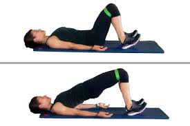
Image result for bridges with therabandBridges with theraband :

Loop a medium band around knees with feet shoulder width apart. LIft rear off mat while pushing knees outward toward band. Hold and slowly return.

Ball at the wall squat: By placing an exercise ball between you and a wall, a standard squat position becomes a wall squat. Wall squats focus on working the lower body and are great for working the quadriceps (front of the legs) and the butt.

Image result for wall squat with ball


Crab walk: 

Begin by sitting on the floor with your feet hip-distance apart in front of you and your arms behind your back with fingers facing hips. Lift hips off the floor and tighten your abs. Start “walking” forward by moving your left hand followed by your right foot; and then your right hand followed by your left foot. Walk four or more steps as space allows, then walk back. Continue back and forth for desired amount of time

Monster walk


Image result for monster walk

Monday, 4 March 2019

Cervical Radiculopathy

Radiculopathy can be quite painful. The condition is commonly referred to as a pinched nerve, with pain, weakness or numbness spreading down the arm.The term radiculopathy comes from radix = “root”, with the nerve root being the common site of nerve pinching. Because this compression, you can feel the pain through the nerve pathway according to the nerve roots involved.


Causes:

A radiculopathy generally occurs with irritation of the nerve root as it exits the spinal column. This irritation can be due to a number of reasons including:

  • physical compression of the nerve from a bulging disc or bony growth (osteophyte) at the edge of the vertebra, tumour, fracture . etc
  • chemical irritation from swelling around the nerve root .

Symptoms of a Cervical Radiculopathy:

Pain is the most common symptom, with the worst pain often being felt further away down the arm.
Commonly  pain referral patterns in accordance with compression of nerves at different levels of the cervical spine.
Compression of the nerve may also lead to impaired nerve function, meaning that you made have areas of reduced sensation in the arm, or reduced power in muscles supplied by the compressed nerve.

Diagnosis:

Your physiotherapist is skilled at determining which structures may be causing your neck and arm pain. There are certain questions that they may ask you to help narrow down the diagnosis and this helps to guide a hands-on assessment. Your physio will palpate the muscles and joints around your neck, as well as perform testing of the nerves.If further investigation is required an MRI is the best way to diagnose a cervical radiculopathy, however, a CT scan or X-ray can still help with diagnosis.

Cervical Radiculopathy Treatment:

Pain Relief. Minimise Swelling & Injury Protection ( Phase 1)

Managing your pain. Pain is the main reason that you seek treatment for this condition.
Managing your inflammation. Inflammation it best eased via ice therapy and techniques or exercises that deload the inflammed structures.
Your physiotherapist will use an array of treatment tools to reduce your pain and inflammation. These include: ice, electrotherapy, soft tissue massage, joint mobility techniques and use of taping techniques or an arm sling to off-load the injured structures.

Restoring Normal ROM & Posture (Phase II)

Once  your pain and inflammation settles, your physiotherapist will turn their attention to restoring your normal joint range of motion (ROM), muscle length, neural tissue mobility and resting muscle tension.
Treatment may include joint mobilisation and alignment techniques, massage, muscle stretches and neurodynamic exercises, plus acupuncture, trigger point therapy or dry needling. Your physiotherapist is an expert in the techniques that will work best for you.

Restore Normal Muscle Control & Strength (PHASE III)

Your physiotherapist will assess your muscle recruitment pattern and prescribe the best exercises for you specific to your needs.
Physiotherapists has developed their own rehabilitation programme to assist their patients to regain normal muscle control of the neck and shoulders. Please ask your physio for their advice.

Restoring Full Function (PHASE IV)

During this stage of your rehabilitation is aimed at returning you to your desired activities. Everyone has different demands for their bodies that will determine what specific treatment goals you need to achieve. For some, it may simply be to walk around the block. Others may wish to do boxing classes or return to a labour-intensive work.

Your physiotherapist will tailor your rehabilitation to help you achieve your own functional goals.

Preventing a Recurrence (PHASE V )

Injury and the pain associated does have a tendency to return. The main reason it is thought to recur is due to insufficient rehabilitation.Your physiotherapist is an expert at identifying underlying joint restrictions and poor muscle patterning, and can help you to work on these to prevent recurrence.

Expecting Results :

Disc and nerve injuries can take a while to recover as the blood flow to these structures can be poor, meaning healing takes longer. Generally, an acute radiculopathy will feel much better in one to two weeks, with resolution by three months. Some cases will take longer, with slower improvements for up to six to twelve months.